
Normal Pressure Hydrocephalus Care
Treating Normal Pressure Hydrocephalus (NPH)
The treatment of choice for NPH patients who show a positive response to diagnostic testing is the placement of a cerebrospinal fluid (CSF) shunt.
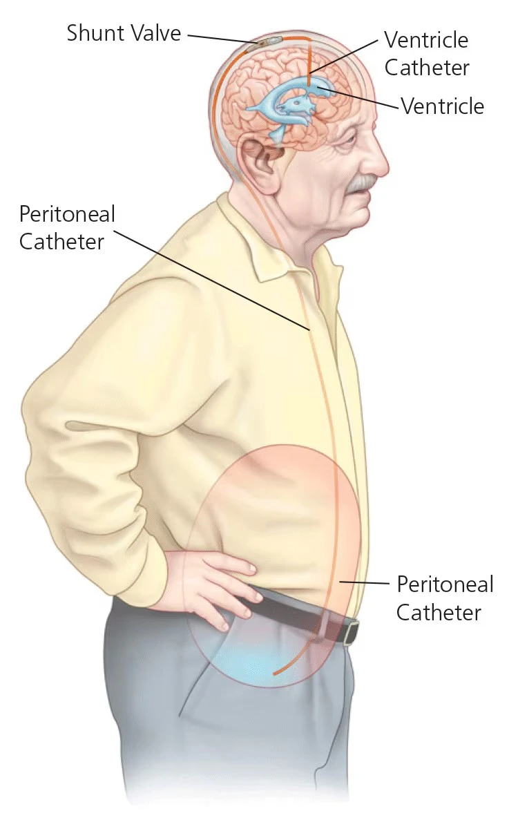
A shunt is an implantable device designed to drain CSF fluid away from the brain thereby, allowing the enlarged ventricles to return to a normal state. As CSF fluid builds and the pressure in the ventricle increases, a one-way valve in the shunt opens, and the excess CSF fluid drains into the abdomen through a catheter, where it is easily absorbed. This technique is very effective in improving the troubling symptoms of NPH.
With a traditional fixed pressure shunt, choice of the correct pressure setting is very important as under-drainage will not improve symptoms, whereas over-drainage can cause symptoms in itself, or predispose to problems such as subdural hematoma. Replacement of a fixed pressure shunt requires removal of the original shunt and implanting a different one.
Surgical revisions such as this can be avoided if your neurosurgeon uses programmable shunt technology. With a programmable shunt, the pressure setting can be adjusted with a special magnetic programmer in your doctor's office, eliminating the need for additional surgery if the initial setting proves not to help, or changes need to be made.

What Is a Shunt?
Shunt systems come in a variety of models but always have two similar components: a catheter, the tubing that transports and diverts the CSF from the ventricles to either the abdominal cavity or right atrium, and a shunt that regulates the pressure or flow of CSF from the ventricles. Valves are manufactured to operate at a specific pressure range. A surgeon chooses a pressure range for the shunt based on experience and the needs of the patient.
Many shunt systems also have a flexible flushing chamber called a reservoir. The reservoir may be housed within the shunt system or added as a component along with the shunt system. The reservoir serves several important functions. It permits the doctor to remove samples of CSF for testing, using a needle and syringe. The doctor also may inject fluid into the shunt system to test for flow to be sure the system is functioning.
The parts of a shunt system are named according to where they are implanted (placed) in the body. The portion of the tube which is inserted into the ventricles is called the ventricular catheter. The peritoneal catheter is the portion of the tube that drains CSF into the abdominal or peritoneal cavity.
If a drainage tube is placed into the right atrium of the heart, it is called the atrial catheter. To get a better understanding of what a shunt system looks like, ask your doctor or nurse to show you samples of the shunts they use. All of the components of a shunt system are made from materials which are well known to be tolerated by the body. For this reason, the entire shunt system is implanted under the skin. There are no external parts.
Fixed vs. Programmable Shunts
When treating hydrocephalus with a shunt system, the choice between a fixed pressure valve and an adjustable (programmable) valve determines how cerebrospinal fluid (CSF) drainage pressure is regulated and whether the setting can be changed after implantation without additional surgery.
If the pressure setting of a fixed pressure shunt proves to be a mismatch after surgery, causing underdrainage or overdrainage complications, the patient must undergo a complete or partial shunt revision, sometimes more than once. This is a limitation of all fixed pressure shunts.
CODMAN® Programmable Shunts
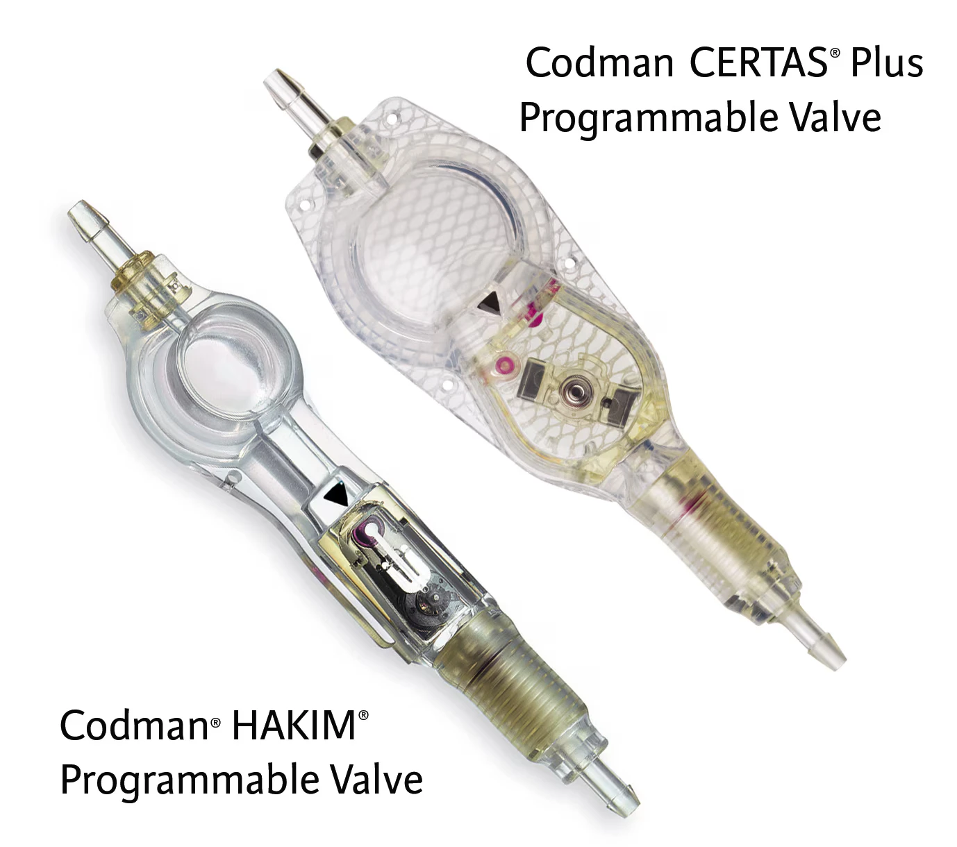
The Codman® HAKIM® Programmable Valve (CHPV) is an example of a programmable valve. It has 18 pressure settings that a surgeon can adjust non-invasively to optimize the treatment.
The Codman® CERTAS® Plus Programmable Valve is another programmable valve, but with 8 different pressure settings, including a virtual off.
Using an exclusive external programming device, the surgeon selects the initial pressure setting prior to the procedure and can then easily adjust the setting at any time and as many times as necessary without further surgery.
The large range of pressure settings allows the surgeon to make very fine adjustments to the pressure in order to get the best resolution of symptoms after the shunt is implanted. The totally non-invasive adjustments take only seconds and can be done right in the office with little or no patient discomfort.
Programming Device
The device used to adjust the pressure setting of a shunt is simply called a programmer. The programmer, used specifically for CHPV, includes an electrical box connected to a transmitter head. Using the transmitter head the shunt is programmed to a certain pressure chosen by the surgeon prior to being implanted. Upon pushing a button, the shunt is changed to the selected pressure in seconds.
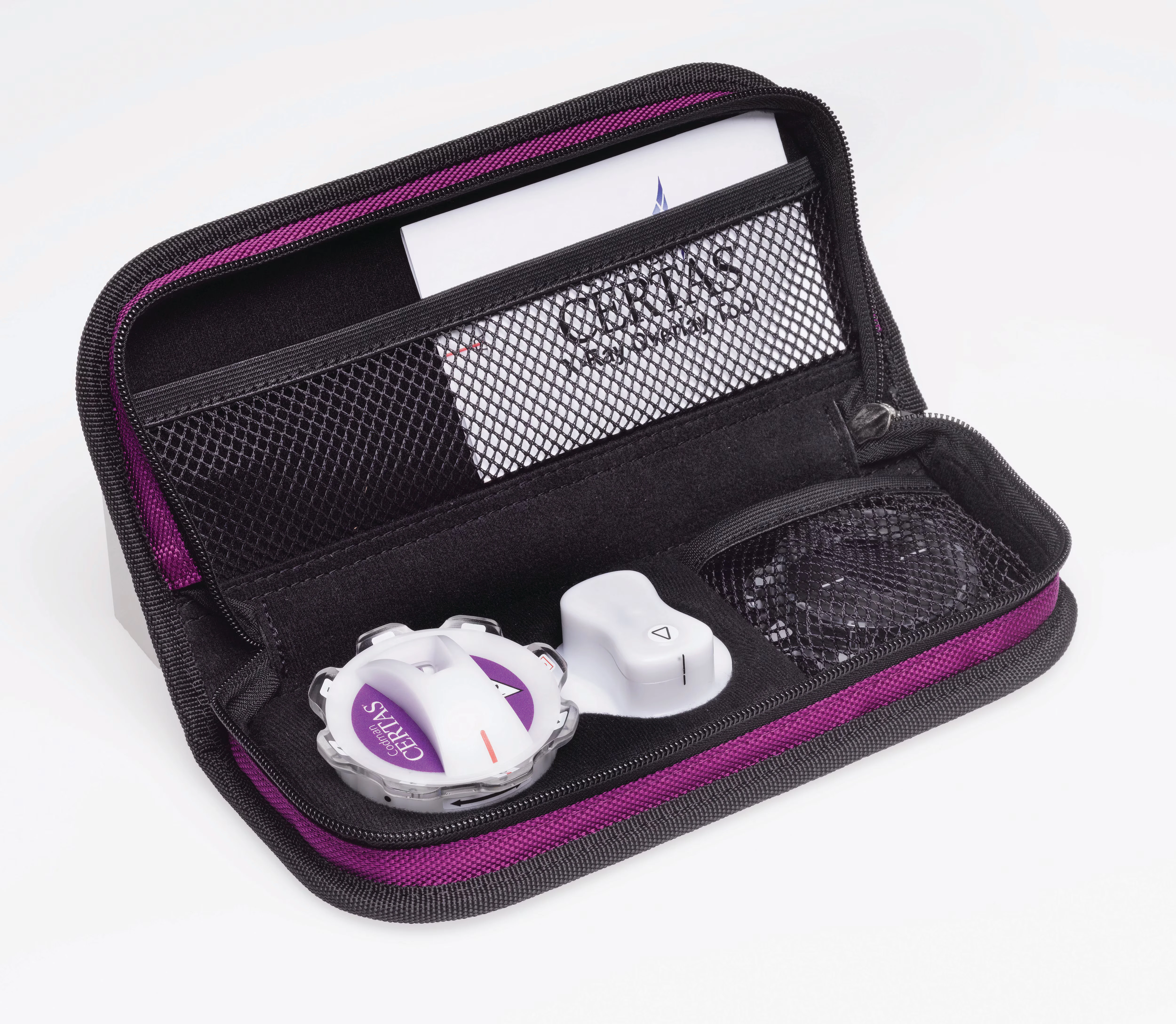
To adjust the setting of Certas Plus Programmable valve, the doctor will use CODMAN CERTAS® Tool Kit. In order to program the Certas Plus valve, the doctor will gently feel the skin to center the locator tool over the implanted valve.
The doctor will then set the indicator tool into the locator tool in order to read the current valve setting. To adjust the setting, the doctor will remove the indicator tool and set the adjustment tool into the locator tool. By simply turning the adjustment tool, the doctor can adjust the pressure settings to the desired performance level of the valve.
- Codman Certas is a registered trademark of Integra LifeSciences Corporation or its subsidiaries in the United States and/or other countries.
- Certas is a trademark of Integra LifeSciences Corporation or its subsidiaries.
- Hakim is a registered trademark of Hakim USA, LLC and is used under license.
Shunt Surgery
The surgical procedure to implant a VP (ventricular peritoneal) shunt usually requires less than an hour in the operating room. After the patient is placed under general anesthesia, their scalp is shaved and the patient is scrubbed with an antiseptic from the scalp to the abdominal area.
These steps are taken in order to reduce the chances of an infection. Incisions are then made on the head and in the abdomen to allow the neurosurgeon to pass the shunt's tubing through the fatty tissue just under the skin. A small hole is made in the skull, opening the membranes between the skull and brain to allow the ventricular end of the shunt catheter to be passed through the brain and into the lateral ventricle.
The abdominal (peritoneal) catheter is passed into the abdominal cavity through a small opening in the lining of the abdomen where the excess CSF will eventually be absorbed. The incisions are then closed and sterile bandages are applied.
After Shunt Surgery
The patient generally stays under careful neurological observation for the first 24 hours following the procedure. Some neurosurgeons prefer to keep the patient flat in bed until nearly all the subdural air introduced during surgery dissipates. The bandages placed on the head and abdomen, covering the incision sites, are monitored for signs of infection.
The patient will generally need to stay in the hospital from three to seven days. Follow-up visits will be necessary to check post-operative status and resolution of symptoms. Additional treatment, such as physical therapy, may be advised to help the patient attain previous levels of motor skills.

Possible Complications
Although shunt surgery is a relatively simple neurosurgical procedure, the decision to undergo insertion of a shunt should not be taken lightly. The treatment of NPH carries greater risks compared to the treatment of children with hydrocephalus, and therefore the operation should be undertaken only if the degree of disability or the progression of the disorder warrants.
The potential complications of shunt surgery should be viewed as those related to the actual operation, plus those that may occur days to years later. A complication can be thought of as any unwanted event related to the surgical procedure itself or the presence of the shunt.
Potential complications may include the infection of the surgical wound or of the CSF (meningitis), bleeding into the brain or ventricles, or a seizure. A shunt infection may be indicated by fever, redness or swelling along the shunt track. Fortunately, these complications are uncommon and can be managed successfully in most cases.
Unlike many other operations in which the surgical risks are highest during the operation itself, most of the common and serious problems associated with shunting can occur weeks or even years after the surgery. The most common problem with shunt systems is that they can become obstructed (clogged). This can occur hours or years after the operation, sometimes multiple times. The likelihood of a shunt obstruction is thought to be about 50% for most patients. For patients with NPH, a shunt obstruction is usually discovered when the original symptoms recur.

Disclaimers
Only your doctor should adjust the pressure setting on programmable valves.
This content provides a summary of the information regarding the appropriate use of the Codman Hakim Precision Fixed Pressure Valve, the Codman CERTAS Plus Programmable Valve, the Codman Hakim Programmable Valve, the Integra OSV II and Low-Flow Flow Regulating Valves.
It is not a substitute for a thorough discussion with your doctor regarding the use of any Codman valve for your specific medical condition. If you have any questions or want more information, be sure to discuss your questions with your doctor
For important safety information, visit here.