Treatment Options
What Are Some of the Diagnostic Tools?
- Ultrasound: a device that uses sound waves to outline the structures within the skull. This is similar to scans that pregnant women have to check that their babies are healthy.
- CT Scan (Computerized Tomography): a technique that uses x-rays to image and outline the size of the ventricles.
- MRI (Magnetic Resonance Imaging): a technique that uses radio signals and a magnet to form images of the brain so that the size and shape of the ventricles can be determined.
- CSF Flow Studies: uses dyes or other materials to trace the flow patterns of cerebrospinal fluid (CSF).
- Neuropsychological Test: a series of questions and answers used to determine if there is a loss of brain function due to hydrocephalus.
- CSF tests to predict shunt responsiveness and/or determine shunt pressure include: lumbar puncture, external lumbar drainage, measurement of CSF outflow resistance, intracranial pressure (ICP) monitoring and isotopic cisternography.
Though there is no way to accurately predict a patient’s responsiveness to any intervention, many doctors find that these tests are helpful in determining the likelihood of a positive response. People who have abnormal bleeding tendencies or take medications that affect bleeding should talk with their medical team about any special precautions before invasive procedures.*
*Courtesy of the Hydrocephalus Association
How Is Hydrocephalus Managed?
A surgical procedure may be performed to divert cerebrospinal fluid (CSF) from the ventricles to either the abdominal cavity or to a chamber in the heart known as the right atrium.
Another technique exists to channel the CSF from the lumbar into the peritoneal cavity. By removing the CSF, the pressure in the skull may return to normal.
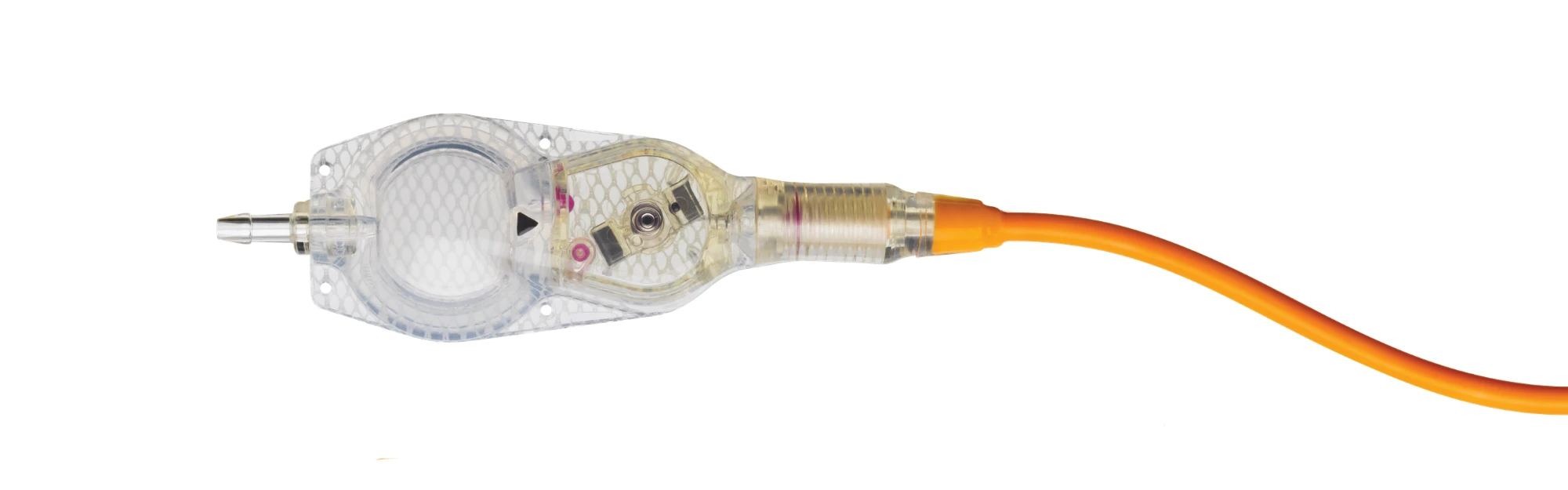
To remove the CSF, the surgeon implants a flexible tube with a valve mechanism called a shunt system. A shunt may help to control the hydrocephalus but is not a cure.